Basic & Translational Research Projects
Bupivacaine Injection Treatment of Strabismus
Strabismus Injection Treatment for Children
| Most strabismus patients are children, and early treatment of infantile strabismus facilitates normal development of stereopsis (depth perception from binocular vision), prevents amblyopia (suppression of vision in one eye), and improves cosmesis. But surgical correction in young children is problematic: [1] additional surgery is often necessary, which is made difficult by scarring from the initial surgery; it would be better if injection treatment were used, at least initially, and [2] strabismus surgery requires prolonged general anesthesia, which can cause enduring cognitive deficits in a developing brain (eg, Rappaport et al 2015).
EOMs lie deep in the orbit, not normally visible, so a technique is needed to accurately place the injection needle within the target muscle. In cooperative adults, electromyography (EMG) signals are recorded from the tip of the injection needle, which is advanced until the relationship of the EMG signal to the patient’s voluntary eye movement indicates proper placement. Children, however, would have to be briefly anesthetized during the injection, and anesthetized muscles show little or no movement-related electrical activity. They will, however, contract in response to electrical stimulation. Thus, we are developing a method to accurately target electrically silent muscles using the tip of the injection needle to stimulate the target muscle, causing contraction and movement of the eye in a characteristic direction. Having demonstrated efficacy in a rabbit model, we now plan to compare stimulation against EMG guidance in adults, documenting injection accuracy using motility measures and MRI examinations of the muscle following injection, as is routine with our adult injection patients. Application in children would follow, under protocols defined by these results. | Reanimating Paralyzed Eye Muscles
| Blepharospasm sufferers may be functionally blind despite having normal eyes, because of spasms in surrounding facial muscles and inability raise their eyelids. The cause is unknown, and may be present from birth or develop later. Botox injection can relieve the spasms but leave patients unable to open their eyes or keep them open. Functional electrical stimulation (FES) of the muscle that raises the eyelid (the levator palpebrae superioris or LPS), could provide these patients with useful vision. Surgical lid elevation is the current treatment, but static repositioning makes normal eye blinking and lid closure problematic. Programmable, coordinated, binocular elevation by FES would be far superior, both functionally and cosmetically.
Similarly, patients suffering from paretic strabismus (misaligned eyes or gaze limitations caused by weak or paralyzed muscles) could be rehabilitated with FES of the extraocular muscles (EOMs). The signal for controlling stimulation of a paretic EOM could be derived from the intact innervation of its antagonist (the opposing muscle in the same eye), with which it normally has a reciprocal relationship. Implantable pulse generators (IPGs), already approved for other applications, are suitable to connect directly to electrodes we have developed for implantation on EOM and LPS.Our aim is to reduce FES of eye muscles to clinical practice. The focus of our work is development of electrodes that are both safe and effective, tested in animals with realistic stimulation regimens. As we develop stimulation parameters, we will refine simultaneity of binocular stimulation and coordination of reciprocal stimulation, and will evaluate tissue tolerance and electrode durability. With these results in hand, we will be in a position to design clinical trials. We’ve already produced useful lid elevation in rabbits as long as a year following implantation, with no evidence of tissue damage. We’ve designed a new electrode for improved reliability, and will also test capacitive electrodes, which may provide even gentler stimulation.
| Oculomotor Unit DiversityExtraocular BiomechanicsDirection Perception & Sensorimotor Adaptation
FabLabDevices developed for our work and made available to our colleagues.
|
| Measuring Physiological Muscle Force
Our "MFTs" are only way to measure physiological eye muscle forces. Learn about:
- An EOM force transducer for intraoperative use in humans.
- A digit flexor tendon force transducer for key-pressing studies during carpal-tunnel release surgery.
- Physiological EOM forces in alert NHPs, and brainstem control of eye movement.
- MFT fabrication & implantation (more than you wanted to know).
|
MethLabTools and techniques developed for our work, but of more general utility.
|
| Orbit™ 1.8 on Intel® (OOI)
Orbit is a unique software tool that provides easy access to a sophisticated biomechanical model, able to simulate classical strabismus syndromes and data from individual cases, clarifying diagnostic and treatment possibilities in well-defined physiologic terms. It is now available in an emulation environment that runs under OSX on modern Intel Macs. |
|
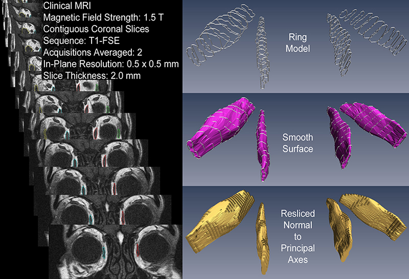
| Analysis of Extraocular Muscle MRI & CT Images
We've developed procedure for quantifying crossectional areas and volumes, which minimizes errors and biases, and which should replace the procedure we introduced in 1989.
|
|
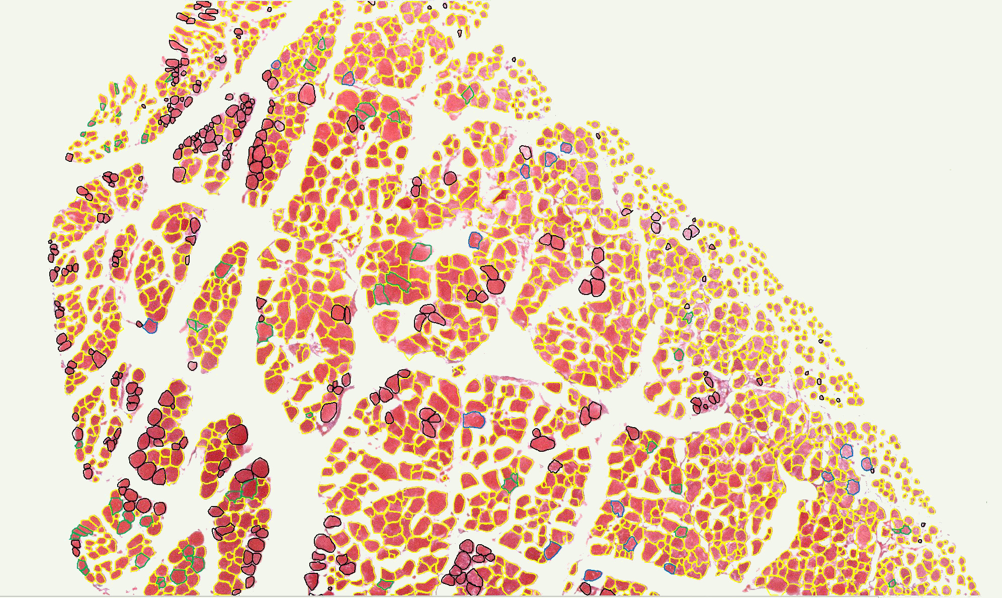
| Segmenting & Quantifying Muscle Fibers in Stained EOM Crossections
Quantitative histology of extraocular muscles (EOMs) has required sampling because of the many thousands of fibers in a crossectional slice, but unfortunately, EOM is highly inhomogeneous making bias difficult or impossible to avoid. We’ve developed a method to segment and measure essentially all the fibers in a section, using an operator-aided automatic process to calculate fiber areas and other statistics, while excluding voids, connective tissues, blood vessels, and nerves. A watershed algorithm segments the histological image based on luminance, and then filters out segments that don’t have shapes and colors characteristic of muscle fibers. Missed fibers are added, grouped fibers are split, and fiber fragments are merged manually, with computer-aided tools (IPP-9.1).
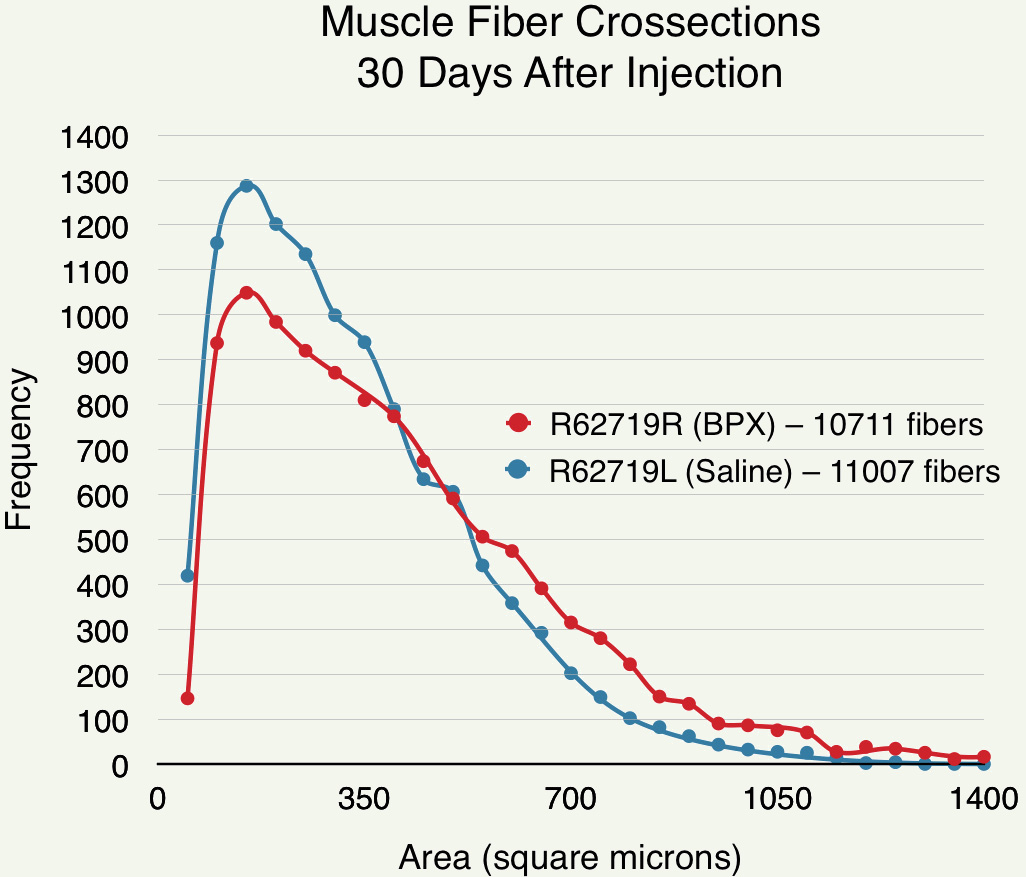
Early results suggest that BPX injection changes the distribution of muscle fiber diameters in favor of larger fibers, clarifying the nature of the overall muscle size changes and alignment corrections observed in humans.
| | |
| EOM Fiber Segmentation. Fibers outlined in yellow were automatically segmented. Fibers in other colors required some manual intervention. With high-quality histological processing, automatic segmentation of about 80% of fibers can be achieved. | Effect of BPX Injection On Muscle Fiber Size. Crossectional areas of all fibers in posterior sections (6 mm anterior to the muscle origin) of rabbit SR muscles 30 days after injection with BPX or saline.
| |
|
| LabOS Data Acquisition & Control for Biomedical Experiments
A software system providing both a rich user interface and deterministic closed-loop control at biological rates of experiments specified by complex protocols. [project stalled!] |
|
|